3D Mammography
Schedule Your Screening MammogramMammograms use low-dose X-rays to examine the breasts and are commonly used to detect breast cancer and other breast changes.
Screening mammograms are performed on women who have no signs or symptoms of breast cancer, as a preventative measure. Diagnostic mammograms are conducted when there is a lump or other concerning sign or symptom –such as unusual breast pain, tenderness, sensitivity, or discharge – or if there is a history of breast cancer.
At WakeMed Imaging, 3D mammography is our standard of care for early detection of breast cancer. It combines multiple breast x-rays to create a 3D picture of the breast, allowing for clearer images and better detection of breast abnormalities, especially in dense breast tissue.
Who Should Get a Screening Mammogram?
The American Cancer Society recommends the following for women at average risk of breast cancer. A provider may recommend an earlier starting age for women with a family history of breast cancer. Early detection makes a critical difference, so starting routine screenings at the right time is essential.
- Women between 40 to 44 have the option to start screening with a mammogram every year.
- Women 45 to 54 should get mammograms every year.
- Women 55 and older can switch to mammogram every other year, or they can choose to continue yearly mammograms. Screening should continue if a woman is in good health and is expected to live at least 10 more years.
Benefits of 3D Mammography
3D breast mammography (also known as breast tomosynthesis) combines multiple breast X-rays to create a three-dimensional picture of the breast. It is the most modern screening and diagnostic tool available for early detection of breast cancer. 3D mammogram machines can quickly create 3D images in one-millimeter slices, as well as standard 2D mammogram images.
3D renderings of breast tissue provide greater visibility for the radiologist and more details about the breast tissue. By combining 3D imaging with standard mammograms, we can reduce the need for follow-up imaging, detect more cancers than 2D screenings, have greater clarity through denser breast tissue and reduce the rate of false positive readings.
PODCAST
WakeMed Voices Podcast explores a variety of health and wellness-focused topics. Hear the answers to your most common questions about mammograms – from when to start getting them to what the results really mean.
What to Expect
Convenient Locations
With locations throughout the Triangle, you can schedule your screening mammogram online or walk-in at your convenience.
A provider referral is not needed to schedule a screening mammogram.
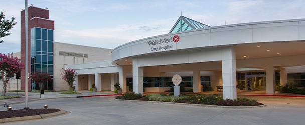
Imaging - Cary Hospital
1900 Kildaire Farm Road - Cary, NC 27518
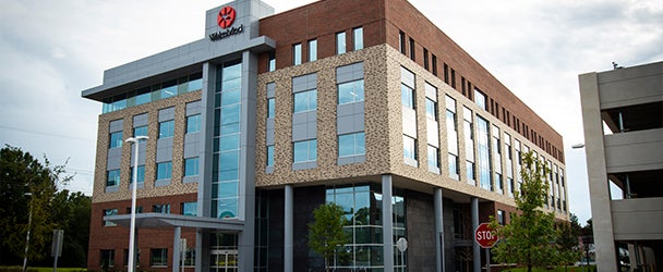
Imaging - Medical Park of Cary
210 Ashville Avenue Suite 130 - Cary, NC 27518
Imaging - North Hospital
10000 Falls of Neuse Road - Raleigh, NC 27614
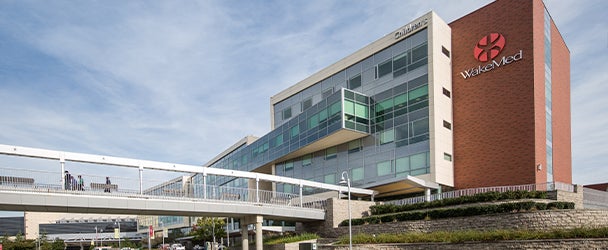
Imaging - Raleigh Campus
3000 New Bern Avenue - Raleigh, NC 27610
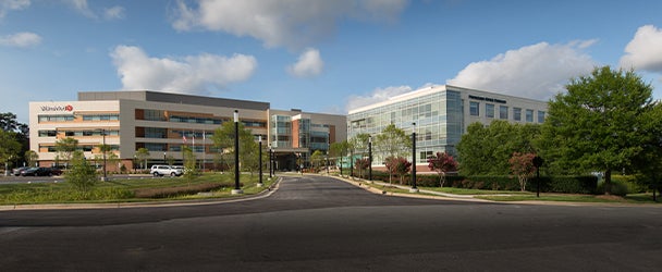
Imaging - Raleigh Medical Park
23 Sunnybrook Road Suite 110 - Raleigh, NC 27610