Save the Tatas: Get a Breast Mammogram
October 28, 2024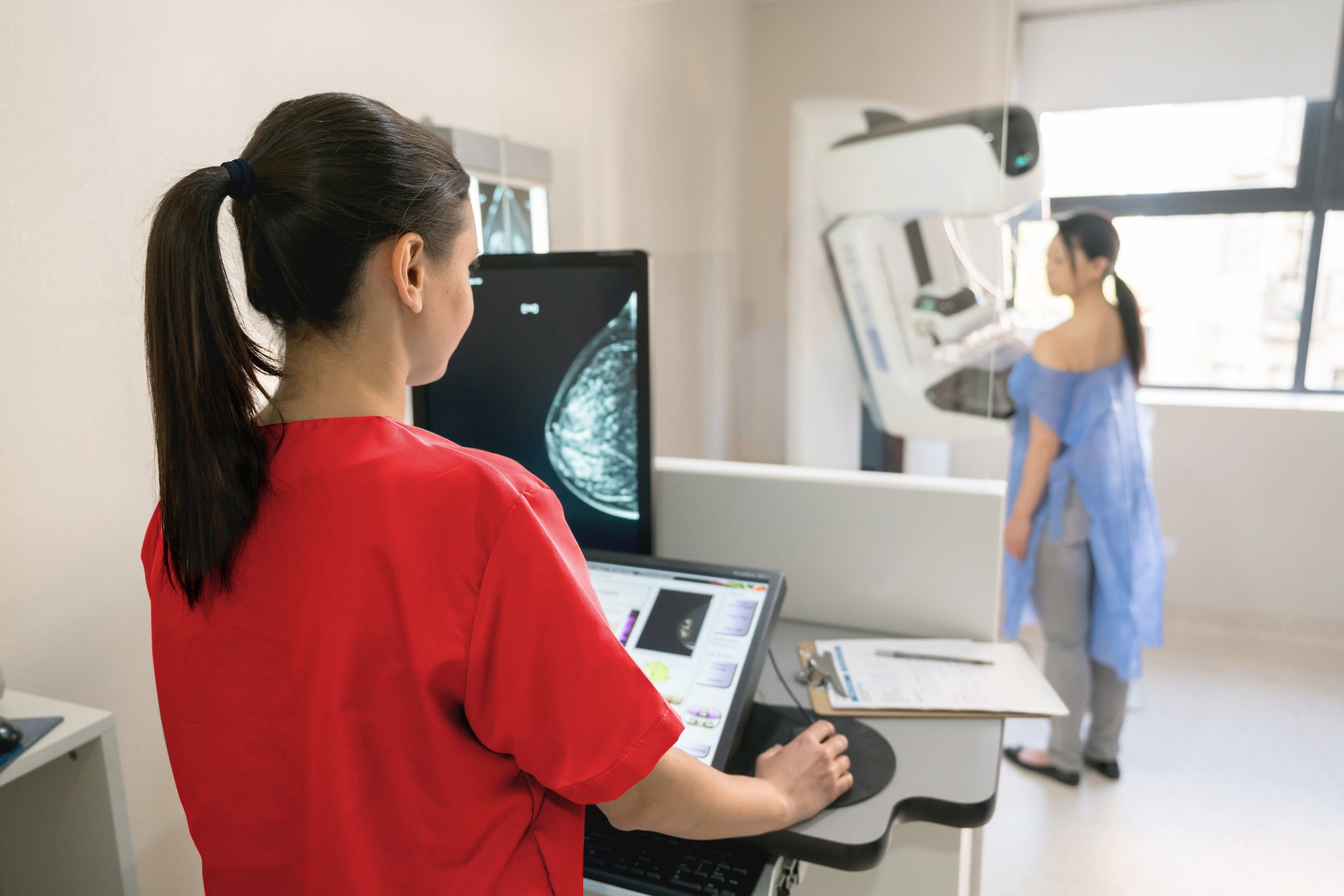
By: WakeMed Health & Hospitals
Categories: Breast Mammography, Cancer Care
Tags: Breast Cancer, Breast Diseases
Breast cancer is the second most common cancer among all women in the United States and is the most common cancer among African American women.
Annually, approximately 128 of every 100,000 women will be diagnosed with breast cancer and about 19 in every 100,000 women will die of breast cancer. Early detection is critical for life-saving care, and the mammogram is the most effective screening tool to identify a potentially malignant tumor at its earliest stage.
With this is mind, we sat down with WakeMed North Hospital imaging manager Dana Knapp, BS, CRA, R.T. (R)(M), to learn more about breast imaging and why it is so important for women to get their annual mammogram.
The Importance of Breast Mammograms
Annual mammograms are very important, and so are monthly self-checks while showering. Early detection is key in beating breast cancer. Women should start mammograms at age 40 and continue through age 74. A provider may recommend an earlier starting age for women with a family history of breast cancer.
Self-checks can be done while standing, showering and lying down. Breast will feel different during menstruation, so it is important to know the differences during different times of the month.
- To self-check while standing, remove the shirt and bra, and use the pads of the three middle fingers to feel breasts in a circular pattern. Start with light pressure, then increase to medium and firm. Check under your areola and squeeze the nipple for discharge.
- When showering, use fingertips to feel breasts in a circular pattern.
- When lying down, breast tissue spreads out, making it easier to feel lumps.
When to Start Breast Mammograms
The American College of Radiology (ACR) suggests that women at average risk for breast cancer begin yearly breast cancer screenings between the ages of 40 and 44. Women 45 to 54 should schedule a yearly mammogram, and women 55 and older may elect to stick with their yearly schedule or switch to a mammogram every other year. Women with a family history of breast cancer, should consult their primary care provider or gynecologist who may suggest beginning mammograms as early as age 35 — this will vary depending on whether an immediate relative (mother) had breast cancer versus an extended relative (aunt, grandmother, cousin, etc.) had breast cancer.
Men and transgendered individuals are also at risk for breast cancer. According to Susan G. Komen, there are different screening recommendations for men at higher risk and transgender people.
Breast Mammogram Safety
Unfortunately, misinformation is ongoing when it comes to breast mammography. Some believe that mammograms are not safe and may lead to cancer or hormonal issues, but this is not true. Breast mammograms do not cause cancer. Neither do they impact the functioning of the thyroid. Mammogram radiation is very minimal and exposes women to significantly less radiation than even a chest X-ray. Plus, since it is typically performed once per year — as opposed to on an ongoing basis throughout the year — it is relatively harmless.
Dana Knapp shares, "A mammogram exposes someone to as much radiation as she would receive over about a seven-week period of naturally occuringoccurring exposure to the sun. It is a very minute exposure, and the risk versus benefit makes it clear that mammography is worth it."
Others believe the mammogram is extremely painful. While patients should feel pressure, the mammogram should not cause stinging or burning pain. If this occurs, the technician may need to readjust the patient on the machine. Each picture only takes about 30 seconds to capture, so the pressure does not last long. The benefits of the breast mammogram, undoubtedly, outweigh any potential discomfort.
3D Mammography
At WakeMed Outpatient Imaging locations, we utilize the highest standard equipment for mammography — as do our imaging partners, Raleigh Radiology. All of our machines are three-dimensional (3D) tomosynthesis mammography units manufactured by Hologic. They are FDA-approved and considered the “BMW” of mammography units. Our friends at Raleigh Radiology also offer 3D tomosynthesis scans at each of their locations. The entire procedure takes approximately five to seven minutes.
The 3D mammogram can detect breast cancer before a patient has any symptoms or feels a lump. Studies have shown that 3D mammograms are more effective than 2D mammograms at detecting early-stage breast cancer.
Knapp explains, "Consider the difference between 2D mammography and 3D mammography as seeing the front and back cover of a book, versus flipping through each page of a book. Our 3D mammography goes through every layer of a person's breast, including very dense breast tissue. In fact, 3D mammography is 40 percent more effective in locating cancers than 2D mammography."
Results
At WakeMed, we pride ourselves in delivering fast results. We realize that waiting can be nerve-racking. The faster the primary care physician receives imaging test results, the faster patients can begin treatment and get on the road to recovery. We make every effort to streamline this process.
Knapp notes, "WakeMed is regulated by the FDA, under the Mammography Quality Standards Act (MQSA). We undergo a rigorous accreditation every year to ensure we're meeting these standards. We provide patient results letters, which contain information about the composition of breast tissue and any Breast Imaging Reporting and Data System (BI-RADS) concerns. The minute a screening mammogram is resulted, read and finalized, that letter is generated, printed out and sent, unless we are waiting for outside imaging to compare to. If the radiologist sees a BI-RADS concern, a patient navigator will call the patient to schedule a diagnostic mammogram. If a woman has heterogenous or extremely dense breast, she will be provided guidance for follow-up with her primary care doctor for potentially more screening."
If the mammogram is normal, the referring provider (primary care or OB/GYN) is also notified of these results so s/he can update the patient.
Test results for patients seen at WakeMed Outpatient Imaging will also be shared via the patient’s WakeMed MyChart account.
A Word About Dense Breast Tissue
If a woman with dense breast tissue has a normal reading, she will also receive a notification of these findings.
"Over 50 percent of women over the age of 40 have heterogeneously dense or extremely dense breast tissue," observes Knapp. "The goal of FDA guidelines is to better inform those patients of the steps to take or the choices they have available."
If the mammography report identifies the patient’s breast density as "Heterogeneously dense, which may obscure small masses” or “The breasts are extremely dense, which lowers the sensitivity of mammography,” the lay summary must include the statement:
“Breast tissue can be either dense or not dense. Dense tissue makes it harder to find breast cancer on a mammogram and also raises the risk of developing breast cancer. Your breast tissue is dense. In some people with dense tissue, other imaging tests in addition to a mammogram may help find cancers. Talk to your health care provider about breast density, risks for breast cancer and your individual situation.”
In addition to dense breasts making it harder to detect cancer, women with dense breast tissue are more likely to develop breast cancer though they are not more likely to die of breast cancer.
According to the National Cancer Institute, "Dense breasts are a risk factor for breast cancer. That is, women with dense breasts have a higher risk of breast cancer than women with fatty breasts. This risk is separate from the effect of dense breasts on the ability to read a mammogram."
Women with dense breast tissue may receive guidance from their primary care provider, after doing a full risk assessment, recommending they add breast MRI or ultrasound to their screening plan. WakeMed offers screening breast MRI as well as information on insurance coverage and co-pays.
According to Knapp, "MRI is a great tool to find small cancers, lesions and small calcifications. If a woman has implants along with dense breast, MRI is helpful in locating potential spots since implants can hide cancers. A combination of 3D mammography and MRI are most effective in detecting breast cancer in these individuals."
An Abnormal Reading
If the mammogram results are abnormal, a patient navigator will contact the patient by phone for a follow-up diagnostic mammogram. Since North Carolina is a right-to-know state, all results — including abnormal readings — are shared just as they would be for a normal reading.
Because letters are sent quickly, a woman may receive the results before the care team can call to schedule a diagnostic mammogram. In that case, she is welcome to make that follow-up appointment herself.
Statistically, 12 percent of patients who come in for a mammogram are called back. Of that 12 percent, just one in 10 are diagnosed with breast cancer.
At WakeMed, we are cautious and thorough, so if a woman’s image has a change from her prior mammograms or a spot stands out on an image, she will be called in for a diagnostic exam to rule out any possibility that she has an area that is suspicious. Even a slight difference will result in a call back for additional imaging because we want to make sure a biopsy is not necessary.
Why Choose WakeMed
Patients should turn to WakeMed for all their breast mammography needs because we provide the highest caliber in 3D standard-of-care imaging; ACR accreditation; exceptional care through our top-notch, credentialed team; and a caring, patient-first focus.
At WakeMed, we have the best 3D mammography equipment currently available, and our state-of-the-art mammography machines are available at every location. Our machines are inspected annually per the guidelines of the Mammography Quality Standards Act, so our standards are also of the highest quality.
WakeMed Breast Imaging Services is ACR-accredited in 3D Mammography, Breast Ultrasound and Stereotactic Breast Biopsy. The ACR accreditation is a peer-reviewed, educationally focused evaluation process to help ensure patients receive the highest level of image quality and safety. ACR accreditation is considered the “gold standard” of breast care.
Our expert care team is among the best and brightest. WakeMed breast imaging radiologists are board certified, licensed and solely dedicated in breast imaging. Our certified technologists are compassionate and committed to excellence in their work and care of their patients.
We realize that our team greatly impacts the patient experience. Patients come to get breast mammograms sometimes scared and full of anxiety. An empathetic technologist can make all the difference. Our goal is always to work collaboratively as a team to ease anxiety and fear from the moment a patient walks through our doors and throughout her entire care experience. And, while we hope that no one ever hears the words, “You have breast cancer,” if a patient is diagnosed, our WakeMed Cancer Care - Hematology & Medical Oncology and Breast Surgery teams are ready and waiting to deliver exceptional and compassionate care.
About Dana J. Knapp, BS, CRA, R.T. (R)(M)
 Dana Knapp is the manager of WakeMed North Hospital's imaging and cardiovascular departments. She also oversees the women’s imaging program at WakeMed. She earned her bachelor’s degree in health care management at Bellevue University in Bellevue, NE. She earned an associates of radiologic technology at New Hampshire Technical Institute in Concord, NH, and an associates of science at Colby Sawyer College in New London, NH. She is also a certified radiology administrator through the American Hospital Radiology Association.
Knapp's longstanding clinical interest has been women’s imaging and early detection of breast cancer. Prior to WakeMed, she managed the women’s imaging program for the University of Colorado Hospital system, implementing 3D as standard of care and developing the first mobile mammography coach offering 3D technology. Knowing the positive impact on patient outcomes, increasing access to mammography, along with education on the importance of screening mammography are her main mission drivers.
Outside of work, Knapp loves to spend time with her husband and four adult children, especially her new granddaughter! She enjoys skiing, golfing and hiking.
About WakeMed Breast Imaging
When it comes to fighting breast cancer, early detection is the best defense. WakeMed Breast Imaging Services offers comprehensive breast imaging and related testing to help physicians find and treat breast cancer at its earliest stage.
WakeMed Raleigh, WakeMed Cary Hospital and WakeMed North Hospital are all designated as Breast Imaging Centers of Excellence locations with the American College of Radiology.
Digital Mammography
Advanced screening and diagnostic mammography are conveniently available in WakeMed locations across Wake and Johnston Counties.
3D Digital mammography decreases the need for additional imaging, detects more breast cancer, and improves detection of breast cancer in dense breast tissue. Breast images are immediately sent to a radiologist and viewed for diagnosis on a high-resolution monitor.
Locations
- WakeMed Cary Hospital
- WakeMed North Hospital
- WakeMed Raleigh Medical Park
- WakeMed Cary – Medical Park of Cary
Imaging locations in partnership with Raleigh Radiology:
Sources
- American College of Radiology
- Susan G Komen
- American Cancer Society