Diagnostics
Time is Brain
Immediately calling 911 for emergency transportation to WakeMed or the closest acute stroke treatment hospital can improve the chances for a stroke victim to preserve as much normal brain function as possible.
Emergency Care
Once at the hospital, WakeMed’s dedicated stroke team — neurologists, neurosurgeons, radiologists, interventional cardiologists, emergency medicine physicians, nurses and therapists — work together to provide advanced, timely stroke diagnosis and treatment.
Various diagnostics are used to determine the location and impact of stroke, these include carotid ultrasound testing, CT imaging, CTA imaging, CTM imaging and MRI, among others.
CT Imaging
Computed tomography (CT) imaging is used to quickly identify or rule out bleeding in the brain. MRI offers precise diagnosis but takes much longer than CT and presents some logistical obstacles that can be cumbersome in the setting of acute stroke, where every minute counts.
CTA Imaging
Computed Tomography Angiography (CTA) imaging uses an injection of contrast material that goes into your blood vessels. It produces pictures of the major blood vessels in your body to help diagnose various conditions, such as stroke.
CTP Imaging
Computed tomography perfusion (CTP), a noninvasive medical imaging technique, provides critical insights into cerebral blood flow, facilitating the timely diagnosis and management of conditions, such as stroke, tumors and other neurological disorders.
2D Echo
This imaging is used to evaluate your heart for potential risk factors for stroke, such as patent foramen ovale (PFO), heart valve disease or the presence of a clot in the heart.
This may also be referred to as a bubble study.
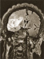
MRI
Magnetic Resonance Imaging (MRI) produces high-resolution images of the body's organs, ligaments, tendons, muscles, and soft tissue with the use of a magnetic field and radio waves.
Carotid Ultrasound

Whoosh! That sound may be telling your doctor that you have carotid artery disease. The carotid arteries carry oxygen-rich blood to your brain, face, scalp and neck. If they narrow or become blocked because of a buildup of plaque (a substance made up of fat, cholesterol and calcium, among other things, found in the blood), your doctor may hear a whooshing sound called a bruit when he or she listens to your carotid arteries through a stethoscope. A bruit is the sound the blood makes when it is flowing, or attempting to flow past an obstacle, such as plaque buildup. When its path is clear, the blood flows quietly and evenly.
Carotid Artery Disease: A Silent Killer
Because it can block blood flow to the brain, carotid artery disease is a leading cause of stroke.
Carotid artery disease can be elusive. People often don't realize they have it until they experience a stroke.
In many cases, we can diagnose carotid artery disease and treat it before a stroke occurs. If your doctor suspects you have carotid artery disease, the next step is typically a carotid ultrasound.
Pain-Free Diagnosis
Carotid ultrasound testing is completely painless. The test uses a high-frequency sound to create images of the insides of two large arteries in your neck, so a cardiologist or vascular surgeon can review their structure and see any plaque buildup. Two ultrasound tests are performed simultaneously: The standard ultrasound provides information about the arteries’ structure, and the Doppler ultrasound focuses on blood flow.
Treatment for Carotid Artery Disease
Treatment for carotid artery disease varies, depending on how far the disease has progressed. Medications such as blood thinners are often prescribed to prevent clotting and stroke. Surgical procedures, including carotid endarterectomy and carotid angioplasty with stenting, are common treatments for those who have advanced disease, and typically are used for patients whose arteries are at least 75% blocked.
Has your doctor listened to your carotid arteries lately? If not, schedule an appointment with your doctor today.