Diagnostics
Time is Brain
Immediately calling 911 for emergency transportation to WakeMed or the closest acute stroke treatment hospital can improve the chances for a stroke victim to preserve as much normal brain function as possible.
Emergency Care
Once at the hospital, WakeMed’s dedicated stroke team — neurologists, neurosurgeons, radiologists, interventional cardiologists, emergency medicine physicians, nurses and therapists — work together to provide advanced, timely stroke diagnosis and treatment.
Various diagnostis are used to determine the location and impact of stroke, these include carotid ultrasound testing, CT imaging, CTA imaging, CTM imaging and MRI, among others.
CT Imaging
Computed tomography (CT) imaging is used to quickly identify or rule out bleeding in the brain. MRI offers precise diagnosis but takes much longer than CT and presents some logistical obstacles that can be cumbersome in the setting of acute stroke, where every minute counts. Learn what to expect from a cranial CT scan.
CTA Imaging
Computed Tomography Angiography (CTA) imaging uses an injection of contrast material that goes into your blood vessels. It produces pictures of the major blood vessels in your body to help diagnose various conditions, such as stroke.
CTM Imaging
Computed Tomoraphy Myelography (CTM) imaging helps to locate conditions that affect the spinal cord and nerves inside the spinal canal. These conditions include infections, spinal stenosis or tumors.
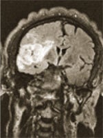
MRI
Magnetic Resonance Imaging (MRI) produces high-resolution images of the body's organs, ligaments, tendons, muscles, and soft tissue with the use of a magnetic field and radio waves.