Preparing for a Mammogram
 What is Mammography?
Schedule Your Screening Mammogram
Mammography is a specific type of imaging that uses a low dose X-ray for examination of the breasts. The images of the breasts can be viewed on film or on a digital mammography workstation. Most medical experts agree that successful treatment of breast cancer is often linked to early diagnosis. Some WakeMed locations also now offer 3D mammography, offering an even more detailed view of the tissue from all angles.
Mammography plays a central part in early detection of breast cancers because it shows changes in the breast up to two years before a patient or physician can feel them. Current guidelines from the U.S. Department of Health and Human Services (HHS), the American Cancer Society (ACS), the American Medical Association (AMA) and the American College of Radiology (ACR) recommend a screening mammography every year for women, beginning at age 40.
The National Cancer Institute (NCI) adds that women who have had breast cancer and those who are at increased risk due to a family history should seek expert medical advice about beginning screenings before age 40 as well as the frequency of screening.
Initial mammographic images are not always enough to determine the existence of a benign or malignant disease with certainty. If finding a spot seems suspicious, your radiologist may recommend further diagnostic studies.
How Should I Prepare for the Procedure?
Before scheduling a mammogram, the ACS and other specialty organizations recommend that you discuss any new findings or problems in your breasts with your doctor. In addition, inform your doctor of any prior surgeries, hormone use, and family or personal history of breast cancer.
Do not schedule your mammogram for the week before your menstrual period if your breasts are tender during this time. The best time to schedule a mammogram is one week following your period. Always inform your doctor or X-ray technologist if there is any possibility that you are pregnant.
The ACS also recommends:
- Do not wear deodorant, talcum powder or lotion under your arms or on your breasts on the day of the exam. These can appear on the X-ray film as calcium spots.
- Describe any breast symptoms or problems to the technologist performing the exam.
- If possible, obtain prior mammograms and make them available to the radiologist at the time of the current exam.
- Ask when your results will be available; do not assume the results are normal if you do not hear from your doctor or the mammography facility.
In addition, before the examination, you will be asked to remove all jewelry and clothing above the waist and you will be given a gown of loose-fitting material that opens in the front.
How Is the Procedure Performed?
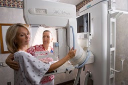
During mammography, a specially qualified radiologic technologist will position you to image your breast. The breast is first placed on a special platform and compressed with a paddle (often made of clear Plexiglas or other plastic).
Breast compression is necessary in order to:
- Even out breast thickness so that all of the tissue can be visualized
- Spread out the tissue so that small abnormalities won't be obscured by overlying breast tissue
- Allow the use of a lower X-ray dose
- Hold the breast still to eliminate blurring of the image caused by motion
- Reduce X-ray scatter to increase sharpness of picture
The technologist will go behind a glass shield while making the X-ray exposure, which will send a beam of X-rays through the breast to the cassette behind the platform and exposing the plate.
You will be asked to change positions slightly between images. The routine views are a top-to-bottom view and a side view. The process is repeated for the other breast.
The examination process should take about half an hour. When the mammography is completed you will be asked to wait until the technologist examines the images to determine if more are needed.
What Will I Experience During the Procedure?
You will feel pressure on the breast as it is squeezed by the compressor. Some women with sensitive breasts may experience discomfort. If this is the case, schedule the procedure when your breasts are least tender. The technologist will apply compression in gradations. Be sure to inform the technologist if pain occurs as compression is increased. If discomfort is significant, less compression will be used.
Who Interprets the Results and How Do I Get Them?
A radiologist, who is a physician experienced in mammography and other X-ray examinations, will analyze the images, describe any abnormalities, and suggest a likely diagnosis. The report will be dictated by the radiologist, and then sent to your referring physician. You will also be notified of the results by the mammography facility. This notification is usually mailed a few days after the official report goes to your doctor.
What Are the Benefits vs. Risks?
Benefits
Imaging of the breast improves a physician's ability to detect small tumors. When cancers are small, the woman has more treatment options and a cure is more likely.
The use of screening mammography increases the detection of small abnormal tissue growths confined to the milk ducts in the breast, called ductal carcinoma in situ (DCIS). These early tumors cannot harm patients if they are removed at this stage, and mammography is the only proven method to reliably detect these tumors.
Risks
The effective radiation dose from a mammogram is about 0.7 mSv, which is about the same as the average person receives from background radiation in three months. The federal mammography guidelines require that each unit be checked by a medical physicist each year to ensure that the unit operates correctly.
False Positive Mammograms. Between five and ten percent of screening mammogram results are abnormal and require more testing (more mammograms, fine needle aspiration, ultrasound or biopsy), and most of the follow-up tests confirm that no cancer was present. It is estimated that a woman who has yearly mammograms between ages 40 and 49 would have about a 30 percent chance of having a false positive mammogram at some point in that decade, and about a seven to eight percent chance of having a breast biopsy within the 10-year period. The estimate for false positive mammograms is about 25 percent for women ages 50 or older.
What Are the Limitations of Mammography?
Interpretations of mammograms can be difficult because a normal breast can appear differently for each woman. Also, the appearance of an image may be compromised if there is powder or salve on the breasts or if you have undergone breast surgery. Because some breasts are hard to visualize, a radiologist may want to compare the image to views from previous examinations. Not all cancers of the breast can be seen on mammography.
Breast implants can also impede accurate mammogram readings because both silicone and saline implants are not transparent on X-rays and can block a clear view of the tissues behind them, especially if the implant has been placed in front of, rather than beneath, the chest muscles. But the NCI says that experienced technologists and radiologists know how to carefully compress the breasts to improve the view without rupturing the implant. When making an appointment for a mammogram, women with implants should ask if the facility uses special techniques designed to accommodate them. Before the mammogram is taken, they should make sure the technologist is experienced in performing mammography on patients with breast implants.
Disclaimer:
Some information from this document is taken from Radiology Info, which is dedicated to providing the highest quality information. However, it is not possible to confirm that this Web site contains complete, up-to-date information. To ensure that this patient education material is accurate, a radiologist has reviewed each section. All information is provided "as is" without express or implied warranty.